The Truth About Radiation in Medical Imaging
If your doctor recommends a CT scan, X-ray, or another imaging test, one of the first questions many patients ask is: “Is radiation in medical imaging safe?”
It’s a reasonable concern. After all, some imaging tests use radiation to create detailed pictures inside the body. But what often gets lost in the conversation is how small these doses are—and how important imaging can be for detecting disease early and guiding treatment.
Understanding the facts about radiation in medical imaging can help put the risks into perspective and give you confidence in your care.
How Radiation Is Used in Medical Imaging
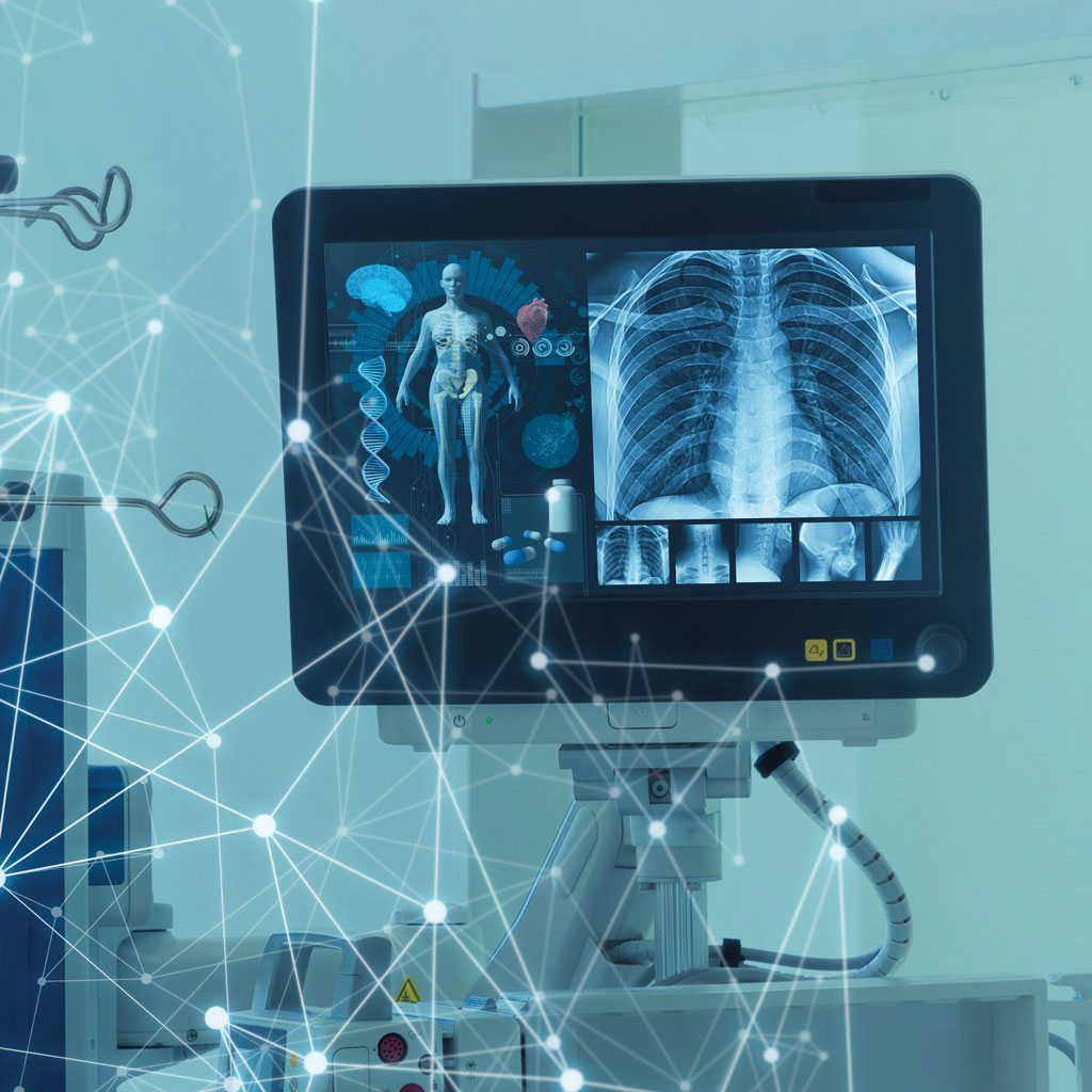
Certain imaging tests use ionizing radiation, which is a form of energy that can pass through the body to create images of bones, organs, and tissues.
Common imaging exams that use radiation include:
These technologies allow doctors to detect fractures, identify internal bleeding, diagnose infections, find tumors, and evaluate organ function—often long before symptoms become severe.
How Much Radiation Comes From Imaging Tests?
Radiation in medical imaging is measured in millisieverts (mSv). For context, everyone is exposed to small amounts of natural radiation every day from the sun, soil, food, and cosmic rays.
In the United States, the average person receives about 3 mSv of natural background radiation each year¹.
Here’s how common imaging tests compare³:
| Imaging Test | Approximate Radiation Dose |
| Extremity X-ray | 0.001 mSv |
| Chest X-ray | ~0.1 mSv |
| Mammogram | ~0.4 mSv |
| CT head | ~2 mSv |
| CT chest | ~7 mSv |
| CT abdomen | ~8–10 mSv |
To put that into perspective:
- One chest X-ray is roughly equal to about 10 days of natural background radiation.
- A CT scan typically falls within 1–10 mSv, depending on the type of scan².
In other words, the radiation from most imaging exams is within the range of what people are naturally exposed to over time.
What About the Risk of Cancer?
Radiation exposure is carefully studied and monitored in medicine. According to the U.S. Food and Drug Administration, the risk of cancer from a single CT scan is considered very small².
For example:
- A CT scan delivering 10 mSv may increase the lifetime risk of fatal cancer by about 1 in 2,000.
- By comparison, the natural lifetime risk of dying from cancer in the U.S. population is about 1 in 5.
That means the additional risk from imaging is tiny compared with the baseline risk everyone already has.
Why Doctors Still Recommend Imaging
While radiation in medical imaging gets a lot of attention, the benefits of imaging are often far greater than the risks.
Imaging tests can:
- Detect cancers earlier, when treatment is most effective
- Identify strokes or internal bleeding within minutes
- Diagnose infections and organ damage quickly
- Help doctors avoid unnecessary surgery
Early detection can literally save lives or prevent serious complications.
For many conditions, imaging provides clarity and peace of mind—either confirming a diagnosis or ruling out something serious.
Modern Imaging Uses the Lowest Possible Radiation
Radiology has made major advances in reducing radiation exposure.
Today’s imaging facilities use safety practices such as:
- Low-dose CT protocols
- Shielding and dose monitoring
- Advanced scanners that optimize radiation automatically
- The ALARA principle (“As Low As Reasonably Achievable”)
These safeguards ensure patients receive the smallest amount of radiation needed to produce accurate images.
Does All Imaging Use Radiation?
No, not all imaging uses radiation.
For example:
- MRI (Magnetic Resonance Imaging) uses magnetic fields and radio waves.
- Ultrasound uses sound waves.
Doctors choose the imaging modality that provides the best information while using the safest approach for your situation.
The Bottom Line: Is Radiation In Medical Imaging Safe?
For the vast majority of patients, yes, medical imaging is considered safe.
The radiation doses used in diagnostic imaging are low, carefully controlled, and monitored by strict safety standards. And the potential benefits—early detection, accurate diagnosis, and peace of mind—almost always outweigh the small theoretical risks.
When it comes to breast health, quality medical imaging is essential for the most accurate results. Bay Imaging Consultants is committed to maintaining the highest standards of quality and professionalism. We are here for you and any of your medical imaging needs.
- Moffatt Cacer Center. https://www.moffitt.org/diagnostic-services/radiology-diagnostic-imaging-and-interventional-radiology/safety-in-radiology/x-ray-safety/
- U.S. Food and Drug Administration. https://www.fda.gov/radiation-emitting-products/medical-x-ray-imaging/what-are-radiation-risks-ct
- Harvard Health. https://www.health.harvard.edu/staying-healthy/do-ct-scans-cause-cancer