What is a CT scan?
A CT, which stands for computed tomography, is based on x-ray technology. A CT scan is a actually a series of multiple x-ray images that are processed by a special computer into thin “slices” of the body’s anatomy. When CT technology was first developed, it was called computed axial tomography, which is why some still refer to the imaging test as a “CAT scan.”
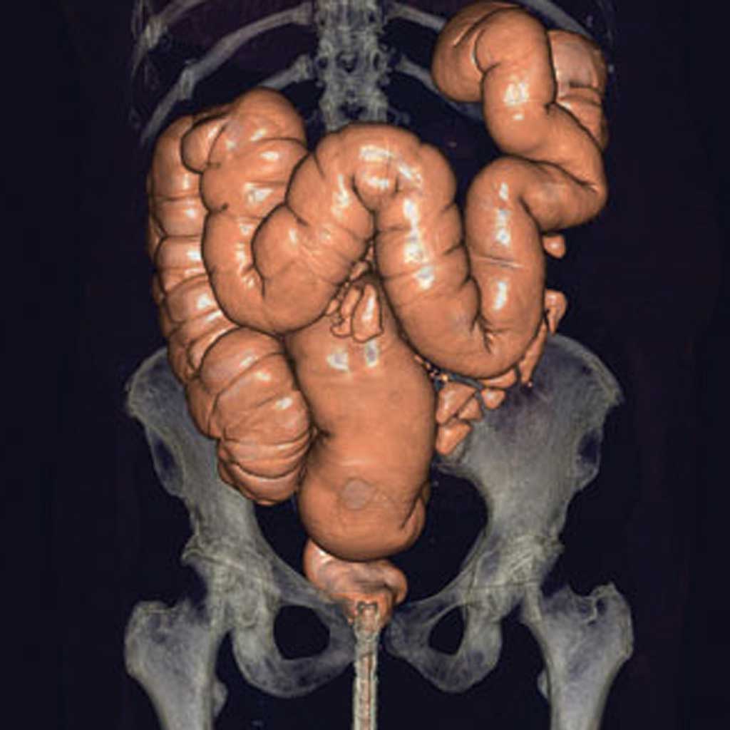
By being able to look at anatomy at various depths, a radiologist gets a more complete view of the inside of the body. This is why CT scans are used to diagnose disease within the body’s organs, identify orthopedic injuries, evaluate the brain and more. CT images can also be reconstructed in three dimensions for a more complete understanding of the nature of the disease or injury, as well as assist in surgical planning.
In some cases, a substance called a contrast agent may need to be injected prior to a CT scan. Contrast is used to help doctors differentiate between certain organs and improve the detection or characterization of suspected diseases. Contrast is also used to evaluate blood flow through the body’s blood vessels. For example, a CT scan of the heart with contrast can show blood flowing through the coronary arteries and help determine if there is a blockage that could lead to a heart attack. This type of CT scan is known as CT angiography (CTA).
CT scans are also used for preventive imaging. Unlike diagnostic CT scans, preventive screening is used to evaluate healthy individuals, proactively looking for disease in its earliest stages. Just like a mammogram is used to find cancer at an earlier, more treatable stage, CT scans can be used to evaluate the body’s organs, such as the heart, lungs, kidneys, brain and other organs to find disease early, giving people the chance to treat their condition it becomes life-threatening.
How does a CT scanner work?
While a conventional x-ray uses a fixed tube that sends the x-rays in a single direction, a CT scanner sends focused x-rays from multiple angles as it rotates around the patient. As the x-rays pass through the patient’s body, they are picked up by a detector (that is also rotating around the patient on the other side) and then transmitted to a special computer and turned into a series of very thin image “slices” of patient anatomy.
These “slices” can be looked at individually by a radiologist or pieced together to form a 3D image of the bone or soft tissue.
Since the 1970s, CT scanners have consistently evolved to achieve better image quality. Newer generations of CT equipment produce smaller and more numerous image “slices,” which is why CT scanner models are named for the number of image slices they obtain. For example, an 8-slice CT captures eight slices of data with each rotation of the scanner around the patient. Modern 64-slice CT scanners generate 64 slices of data with each rotation, which capture thinner images faster. This results in greater image detail as well as faster scan times for the patient.
Modern CT scanners feature an open design, which is not confining or claustrophobic for the patient. The image acquisition speeds are so fast that the scanner can accurately image the heart between individual beats. The cost of CT is lower than other modalities like MRI, which is why CT is often used for an initial diagnosis and to rule out disease or injuries.